Out now: Commentary on infant entrainment study

Our* commentary on Köster et al. (2019) Psych Sci is now available here. Find a TL;DR below.

Entraining rhythms of the brain to study the neurodevelopment of perception and cognition is a fascinating idea. In principle, entrainment endows the experimenter with increased control over the timing of fundamental neural processes. Yet, its appeal has also created a flurry of neuroscientific research that may apply this idea prematurely. Can we simply infer entrainment
just because we are using rhythmic stimulation? It is underappreciated that there is no clear response to this question yet. This should make us more cautious not only in interpreting findings about the role of neural rhythms in perception and cognition, but also in testing rhythmic sensory stimulation as a clinical intervention for psychopathologies.

Can we know? Created with the memer package for R, adapted from a tweet by Matt Craddock.

Köster et al. (2019) studied the neurodevelopment of action expectations by presenting infants with rhythmically flickering stimuli while recording their EEGs. The authors assumed that this rhythmic stimulation entrained endogenous brain oscillations. In turn, they interpreted a band-limited difference between conditions as a modulation of infant theta oscillations. In our commentary, we discuss this assumption critically. We show how a similar pattern of results can arise from a simulated hypothetical scenario. We did not intend to simulate EEG data in their full complexity, but instead present a simplified model that makes a minimal number of assumptions. Crucially, this model does not require the involvement of entrained endogenous brain oscillations. Our findings may serve as a word of caution on inferring entrainment from (conditional effects on) band-limited neural responses in the context of rhythmic sensory stimulation, and interpreting these findings in the context of endogenous neural oscillations, in neurodevelopment and beyond.


Further reads:

Obleser J, Henry MJ, Lakatos P. What do we talk about when we talk about rhythm?. PLoS biology. 2017 Sep 19;15(9):e2002794.

Keitel C, Quigley C, Ruhnau P. Stimulus-driven brain oscillations in the alpha range: entrainment of intrinsic rhythms or frequency-following response?. Journal of Neuroscience. 2014 Jul 30;34(31):10137-40.


* Blogpost based on unpublished article elements produced as part of the submission process. As such, these have been edited by the co-authors Molly Henry, Sarah Jessen, and Jonas Obleser and have been part of the journal review process.

Call for Papers – Rhythms in cognition: re-visiting the evidence

Special issue at the European Journal of Neuroscience now open for submissions.

Guest Editors Christian Keitel (University of Stirling, UK), Manuela Ruzzoli (University of Glasgow, UK), Chris Benwell (University of Dundee, UK), Niko Busch (University of Muenster, Germany), and Laura Dugué (Paris Descartes University, France).

Call for Papers: November 2019
Extended Submission Deadline: December 21, 2020
Online Publication within One Week of Acceptance
Estimated Date of Publication of Final Issue: Spring 2021

Everyday experience may arise from a fundamentally discrete sampling of our sensory environment, just like a movie consists of still frames shown in rapid succession. Already Bishop (1932, Am J Physiol Leg) suggested such a sampling process. He further identified rhythmic brain activity as the phasic neural analogue of a shutter mechanism subserving discrete sampling. Although it contrasts starkly with our intuition of a continuous perceptual flow the sampling idea has been reiterated over the past 90 years. Ever-more precise recordings of human brain activity and novel analysis techniques have seen an unprecedented surge of interest over the last decade. Consequently, we have come to see brain rhythms as the neural implementation of perceptual sampling and the basis of cognitive functions such as attention, memory and language. In an interesting twist however, more recent negative findings on the role of pre-stimulus oscillatory phase on perception suggest that support for discrete sampling as a fundamental mechanism remains equivocal.

In our Special Issue, we call for methodologically principled studies, irrespective of their outcome, to provide us with the most detailed picture to-date as to the conditions under which perceptual sampling, and its consequences for cognition, can (not) be observed. These studies can be original contributions, replication attempts, pre-registered studies or file-drawer experiments that have to follow a thorough methodology and thus allow clear interpretations also of negative findings. We also welcome dedicated reviews, opinion pieces and methodological advances. Studies can address perceptual sampling in vision, audition or other senses by testing its impact on neurophysiological or behavioural performance measures (psychophysics). We encourage authors to make their data openly accessible along with their experimental and analysis codes in order to foster reproducibility and transparency. We invite human studies adopting neuroimaging (EEG, iEEG, MEG, fMRI) and neurostimulation techniques (tES, TMS, sensory entrainment). Animal studies will be a highly welcome supplement.

We are very much looking forward to your submissions to our EJN Special Issue “Rhythms in Cognition: Revisiting the Evidence”.


Flicker-driven brain waves and alpha rhythms – revisited

In our recent study we asked how waves that the brain produces by itself – alpha rhythms – relate to waves triggered by viewing a flickering screen. Both can be of similar frequency (~10Hz, or ten cycles per second) and are easily recorded from the scalp with electrodes (EEG). To recap, we did not find much evidence for a strong link between the two types of brain waves (more info).

However, there are many ways to produce a flicker, and virtually any rhythmic change in a stimulus will likely drive a measurable brain wave. In experiments we typically use rhythmic changes in brightness (light on/off) or contrast (imagine flipping a checkerboard with its mirror image). Less frequently we use changes along other dimensions of visual properties (e.g. colour, motion). But even within a given property dimension there is a lot of wiggle room in the choice of stimulation parameters – some experiments employ low intensity/low contrast stimuli (as we did) others use intense, full contrast checkerboards.

We know that the properties of the flicker can have a profound influence on the shape of the brain waves they are driving. In brief, waves can look more and less sinusoidal depending on stimulation. The best way to visualise this is to look at EEG spectra (- basically a ledger of the sinusoidal oscillations that make up the EEG-recorded waves). If you only see a peak at the flicker rate (say 10 Hz) then the brain wave elicited by the stimulation will be nearly sinusoidal. If other peaks show up at 20, 30 Hz and so on (multiples of 10), then chances are high your waveform looks more rugged.

With all that in mind it is worth considering that different waveforms, produced by different flickers while keeping the frequency constant, may influence the results of an experiment. Looking at our data, is there a possibility that we would have arrived at different conclusions with an alternative flicker approach?

In our experiment we used a relatively unique approach of smooth contrast changes. Put briefly, we presented a slightly changed version of the stimulus on each frame (= one picture of a movie) of the stimulation. A more typical approach is to switch a stimulus on and off repeatedly to arrive at a similar presentation rate. Would this type of flicker produce a similar pattern of results?

I was able to check this in data from an older study. This experiment had a similar set up with flickering stimuli presented on the left (rate = 10.6 Hz) and right (14.2 Hz). Crucially, here stimuli were simply switched on & off to produce the flicker (more info can be found in the original paper). Suffice it to say that (N = 14) participants were shown a cue (left/right) telling them to focus their attention towards the left or right stimulus for the rest of the trial (~3sec). As in our recent study, I tested effects of attention on the power of the intrinsic alpha rhythm and flicker-driven brain waves. To do so I used the scripts available on osf.io/apsyf.

alphaFig

Above, we see a very typical pattern in the scalp map: the power of the alpha rhythm lateralises according to the focus of attention. Focusing on the left stimulus reduces alpha power over the right hemisphere and vice versa (- although the effect seems to be relatively weak for the left hemisphere). Looking at the spectra shows higher alpha power in the 8 – 13 Hz alpha frequency band when participants ignored the contralateral stimulus position. Interestingly, we also see that the stimulation is intense enough to produce power peaks visible in the spectra of what we have called ‘ongoing’ power. Note that these peaks do not necessarily carry the alpha suppression effect (10.6 Hz peak, right hemisphere, purple spectra) and may even show a reversed pattern (14.2 Hz peak, left hemisphere, orange spectra).

SSRFig

Also, the results for the stimulus-driven waves look very similar to our first report: A measure of how well the brain repeatedly tracks the flicker on each side shows clear peaks at the stimulation frequencies 10.6 & 14.2 Hz (and a Harmonic). Both responses increase when participants focus on the respective driving stimulus. Scalp maps give an impression of this effect* across recording electrodes.

To cut it short, despite the differences in stimulation, patterns of results of both experiments are very similar. Thus, different stimulation approaches may produce different waveforms while the effects the experimenter intends to measure on the waveforms can remain comparable (at least in our case, for visuo-spatial attention).


Thanks to Matt Davidson for prompting this re-analysis on twitter.

* Note that these maps look different from the ones published here. This highlights the influence of factors such as stimulus intensity, location and frequency on the variability of how attention effects project to the scalp.

CNS 2019 – Posters

We will be presenting our works at Poster Session F, Tue (26 Mar), 8-10am, at the 2019 CNS meeting in San Francisco.

Go see Anne for Auditory and visual speech perception is predicted by distinct cortical encoding networks [F60]:

mockPost_AK

And stop by at [F130] for Christian’s Spontaneous fluctuations of pupil size and brain rhythms co-vary at rest:mockPost_CK

… or just click on the posters to get the pdf.

Alpha rhythms: Some slow down, some grow stronger

Researchers usually assume alpha brain waves to behave relatively similarly over time. In a new study, led by Chris Benwell and just accepted for publication in NeuroImage, we find that this is not necessarily true for the 1-2 hrs that a typical EEG experiment lasts.

In a re-analysis of data from two previous EEG experiments we saw that alpha underlies two major trends – alpha power increases (an effect previously described) and alpha frequency decreases consistently over time. Both effects maybe related to growing fatigue, the depletion of cognitive resources or a transition from a volitional to a more automatic task performance.

We also found that different sources of alpha rhythms in the brain can show different trends. While some showed both, power increases and frequency decreases, others showed only one if the two trends. Further analysis revealed that both trends do not necessarily depend on each other.


benwellBlog_eff

Figure We usually assume that alpha varies “spontaneously” around a mean and higher alpha leads us to observe a different behaviour. This relationship however could simply be a consequence of both, alpha and behaviour changing “deterministically”, i.e. trending over time. In experiments, we likely observe a “mix” of both sources of variance (also see Benwell et al. 2017 or here).


These findings need to be taken into account when testing for links between brain rhythms (brain state) and behaviour – a link might be reported accidentally just because both, brain state and behaviour change over time – and when attempting to manipulate alpha through stimulation – stimulation frequencies may need to be adapted on the fly, stimulation will need to target a specific alpha generator while leaving others unbothered.


Anne to join University of Dundee Psychology

[1 Jan 2019]

Anne has taken up a position of Lecturer of Psychology at the University of Dundee.

dundee_website

With the beginning of January 2019 she joins the staff at the Psychology Division at the school of Social Sciences. There, she will continue her research into auditory cognition, with a focus on language and speech perception mainly using EEG-based neuroimaging and, this term, teach on individual differences (personality psychology).

Up-and-coming Dundee is one of the sunniest places in Scotland, located on the banks of the river Tay, an hour north of Edinburgh. The University is famous for its inclusive environment.

Flicker-driven brain waves and alpha rhythms

[17 Feb 2019]

Our manuscript Stimulus-driven brain rhythms within the alpha band: The attentional-modulation conundrum has just been accepted for publication in the Journal of Neuroscience. We show that stimulus-driven and intrinsic brain rhythms in the ~10 Hz range (alpha) can be functionally segregated. Briefly put, while one goes up the other one goes down.

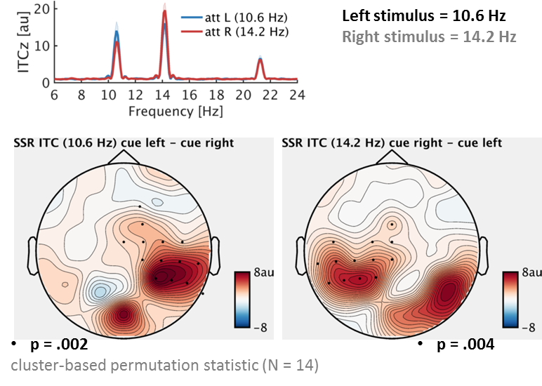
In an experiment, we recorded the brain waves of our participants while they were watching a screen with two stimuli. One, shown on the left, flickered at a rate of 10 Hz and another one, shown on the right, flickered at a rate of 12 Hz. (10 Hz flicker means that the stimulus cycles through a change in appearance or is simply switched on and off 10 times per second.) A very prominent notion has it that this type of visual stimulation is capable of taking possession, or “entrain”, the brain’s intrinsic alpha rhythm. The alpha rhythm can be characterised by its *amplitude* – the difference between peaks and troughs or, bluntly put, how strong it is – and its *phase* – when to expect a peak or trough based on its periodicity. From an entrainment perspective, alpha phase is assumed to lock on and align precisely to the periodicity of the visual stimulation.

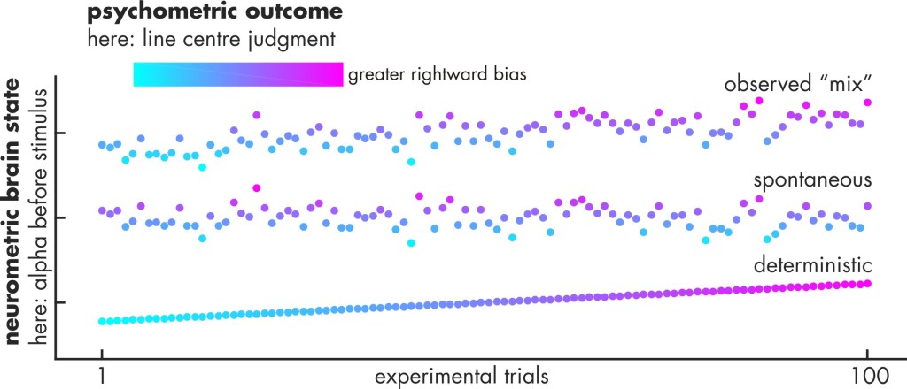
Note that alpha itself has been looked into for almost a century and alpha phase has been tied to another exciting idea: How our brain processes visual input could be more akin to a camera than the continuous make-believe of our daily experience. Hereby, alpha works as a pacemaker that cuts the real-world continuity into perceptual samples or frames just like still frames of a movie. In line with this idea, experiments have shown that we seem to be less sensitive to “see” brief stimuli that pop up during one part of the alpha cycle – in the camera analogy, when the shutter is down – and more sensitive during another part, i.e. when the shutter is open.

Now, the *combination* of perceptual sampling and entrainment puts experimenters in a formidable position to study alpha’s role in perception. It allows them to manipulate alpha phase and exactly time the presentation of stimuli accordingly. Being able to entrain alpha (or other rhythms) through rhythmic visual stimulation would thus be a versatile and easy-to-apply tool – but does it really work?

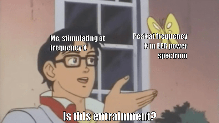
In short, our experiment adds to a line of recent studies that challenge a straightforward alpha entrainment using visual flicker. Our main assumption was this: If it looks like alpha and behaves like alpha, then it should be alpha. *It* refers to the brain waves elicited by watching a 10 Hz flicker. Because the brain response shows up as 10-Hz rhythm in the EEG it does *look* like alpha – especially if you look at it in the frequency domain where it produces a neat 10-Hz spectral peak. “Does it behave like alpha?” we translated into “Does it have the same function?”

One very well documented effect is that alpha power (its strength) shifts according to where we attend to. If we focus our attention somewhere to our left (without actually looking there) then alpha power will go down in our right visual brain – due to the cross-wiring of our visual system from eye to cortex, this is where our left visual world is processed. This alpha decrease works like opening the gates for visual input to venture into further stages of processing. Simultaneously, alpha *increases* in the left visual brain, figuratively closing the gates to unattended, irrelevant sights to our right.

Would a brain response driven by our 10/12 Hz stimulation show a similar effect? If so, that would be strong evidence for a close relationship of spontaneous and stimulus-driven alpha brain waves. Using rhythmic flicker to control alpha experimentally would seem like a readily available manipulation. That was not what we found though. On the contrary – we were able to switch between alpha and the stimulus-driven brain waves using slightly different data analysis approaches. Also, attention had the known suppressive effect on alpha while the corresponding (i.e. same-side) stimulus-driven brain response increased.

These results led us to conclude that we are looking at two concurrent neural phenomena, alpha and flicker-driven brain responses. And each one of them seems to provide us with a different perspective on how attention alters our perception.

Find the specifics and references here.


Note 1: Of course, our results do not rule out alpha entrainment, only that an alpha range stimulus-driven brain wave should not be regarded as sufficient to show alpha entrainment. In the paper (and previous literature) we discuss several, possibly additional conditions that need to be satisfied to give rise to the phenomenon.

Note 2: Data and code to reproduce our results are available here. With minor modifications this code should be applicable to other datasets.


Disclaimer: Views expressed in this digest are mine (CK) and not necessarily shared in all their nuances between the co-authors of the manuscript.

Anne wins Biomag 2018 Young Investigator Award

[28 Aug 2018]

Anne has been one of three Early Career Researchers rewarded with a BIOMAG Young Investigator Award at the 2018 International Conference on Biomagnetism, held in Philadelphia, USA. Nominated as one of 10 finalists she won the award for a presentation of her recent work into the neural foundations of speech comprehension [1].

2018BIOMAG-YIA

The award ceremony took place at the conference gala in the Philadelphia Museum of Art, not only known for its collection of contemporary art, but also for its iconic set of stairs famously featured in the “Rocky” movies.


[1] Keitel A, Gross J, Kayser C (2018) Perceptually relevant speech tracking in auditory and motor regions reflects distinct linguistic features. PLoS Biology ~pdf

First WoRB – Workshop on Rhythms in the Brain – concluded

Last week the first Workshop on Rhythms in the Brain – WoRB – took place here in Glasgow, at the Institute of Neuroscience & Psychology. The organisers thank our speakers Satu Palva, Ayelet Landau, Hartwig Siebner & Niko Busch for their inspiring talks, great discussions and joining our hike up Conic Hill.

Also thanks to everyone who attended the talks, found the time to meet with our guests and our fantastic Admin team, who made it all happen.


news-photo.jpg

Accepted paper: Spontaneous and deterministic fluctuations of pre-stimulus alpha power govern biases in visual line bisection

Chris Benwell’s most recent study shows that an observer’s brain state, just before seeing a transected line, influences their judgment of its centre. Also, these critical brain states do not seem to fluctuate randomly from instant to instant, but trend systematically over time.

Brain states can be defined by rhythmic activity in characteristic frequency bands. We know that the alpha rhythm (roughly ten cycles per second) plays its role in how we take in the visual world around us. For example, strong alpha can shield us from visual impressions. Weak alpha allows for more sensory intake.

Until recently this role of alpha had mostly been shown for briefly presented light flashes just bright enough to be on the verge of being visible. But Chris’ study suggests that alpha may be a gatekeeper for other aspects of visual processing in the brain, too. Here, observers judged whether a line was transected left or right of its centre. Crucially, alpha power before line presentation influenced this judgment.

Line stimuli – Observers had to judge whether lines were interrupted left or right of their actual centre.

More surprising however was that fluctuations in alpha power didn’t come as spontaneous or random as previously thought. Instead, the time observers spent on the task seemed to play a role – at least to some extent. Alpha simply increased over the course of the experiment. And here’s the novel aspect: This deterministic trend predicted a gradual shift in line centre judgments from trial to trial. On average, observers judged the centre to be more to the right than it actually was.

In brief, Chris’ experiment showed that alpha not only influences whether we see very faint stimuli but also how we make judgments about the centre of a visual object. Also, alpha does not just fluctuate randomly but has a deterministic component: When performing a task for an extended period of time alpha inclines gradually. This leads to a sustained and predictable change in our visual perception.

benwellBlog_eff.jpg

Schematic of spontaneous and deterministic influences of alpha (a neurometric) on reported line center (a psychometric) – the colour gradient towards purple indicates that observers showed an increased rightward bias over time meaning that they increasingly judged the line centre more right than it actually was.

These findings beg an interesting question for future research: Can we find other determinants of alpha (i.e. brain state) fluctuations? Moreover, can all ‘spontaneous’ fluctuations ultimately be described by deterministic processes?

The paper has just been accepted for publication in the European Journal of Neuroscience, and can be found here.

Reference
Benwell CSY, Keitel C, Harvey M, Gross J, Thut G (accepted) Trial-by-trial co-variation of pre-stimulus EEG alpha power and visuospatial bias reflects a mixture of stochastic and deterministic effects. European Journal of Neuroscience

This paper is part of the EJN special issue “Neural Oscillations”.